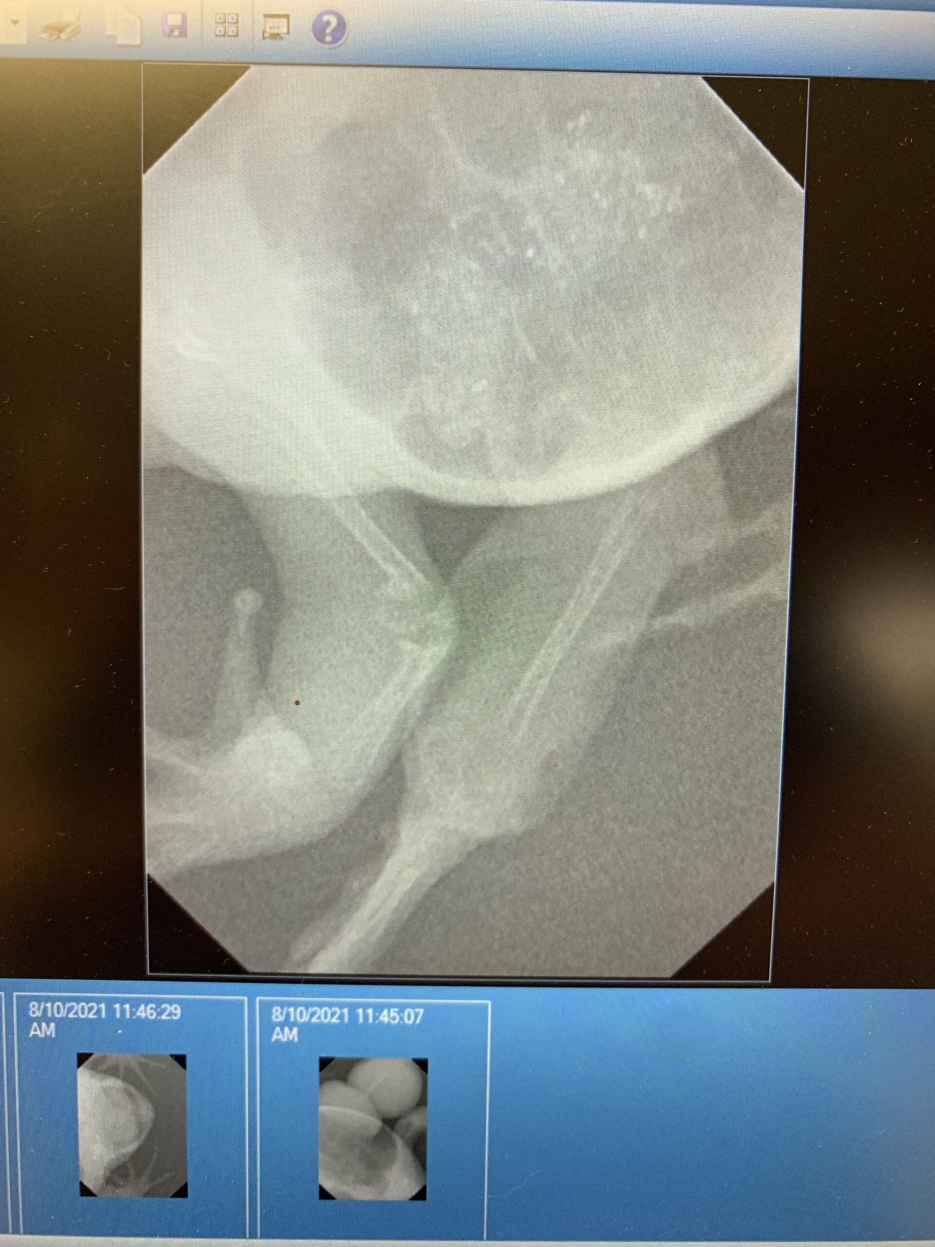
Exotic Diagnostic Imaging
What are the various forms of diagnostic imaging available? There are several options at our disposal. The most common is X-rays, providing a 2D static image of your pet. Ultrasound, also 2D but dynamic, can be directed to different areas. We utilize CT scans, generating a 3D image, and MRI, which excels in examining neurological tissues. Additionally, dental X-rays offer detailed views of small areas, while nuclear scintigraphy assesses organ functionality.
Dr. Stephanie Munyon
Brook-Falls Veterinary Hospital.
What aspects does diagnostic imaging help assess in exotic pets? We utilize imaging to explore aspects not visible during a physical exam. For instance, in rabbits and guinea pigs, we often need clearer views of their teeth due to their small mouths. Imaging allows examination of nasal passages, lung health in turtles, and internal structures in hedgehogs, among other things.
Is sedation necessary for diagnostic imaging in exotic pets? The need for sedation varies depending on the pet. Some may cooperate well, while others may become stressed or too active to remain still. Generally, CT scans require heavy sedation or anesthesia to ensure accurate imaging.
When should X-rays be used compared to MRI, ultrasound, or CT scans for exotic species? X-rays and ultrasound are often the initial choices due to their speed and convenience, especially during appointments. CT scans offer enhanced detail, eliminating superimposition issues associated with 2D imaging. This 3D modality provides clearer interpretation of complex structures.
For further inquiries, feel free to contact us directly at (262) 781-5277 or via email at [email protected]. We aim to respond promptly. Don’t forget to connect with us on social media platforms like Facebook and Instagram.
Explore Our Complete List of Veterinary Services in Menomonee Falls
- Grooming Services
- Wellness Plans
- CT Scan
- Rabbits
- Guinea Pigs
- Ferrets
- Exotic Boarding
- Wing Clipping
- Bird Wellness Appointments
- Bird Nutrition
- Bearded Dragons
- Bird Identification
- Dog Spay & Neuter
- Dog Surgery
- Dog Senior Wellness
- Puppy Care
- Dog Microchipping
- Dog Laparoscopic Surgery
- Dog Flea & Tick
- Dog Diagnostic Imaging
- Dog Dental
- Kitten Care
- Kitten Behavior & Socialization
- Cat Vaccines
- Cat Surgery
- Cat Spay & Neuter
- Cat Nutrition
- Cat Heartworm
- Cat Flea & Tick
- Cat Diagnostic Imaging
- Cat Intestinal Parasites
- Cat Dental
- Pharmacy
- Dental Care
- Surgery
- Laser Therapy
- Advanced Diagnostic Imaging for Pets
